3p Neuronal Imaging
Problems
The in vivo neural activity of living organisms is not easy to be detected,, especially when going deep and maintaining subcellular resolution and signal to noise, in order to extract information about both the structural and functional parts of the brain. With three-photon fluorescence microscopy (3PFM) it is possible to go deeper and therefore create an in vivo scanning microscope to analyze areas of the brain previously inaccessible with this resolution detail.
Technology
In three photons microscopy technique, lasers must emit push into the infrared frequencies and with peak energy levels high enough to enabling a three-photon transition (otherwise very weak). Longer excitation wavelengths reduce the tissue scattering and the higher-order nonlinear excitation increases the background suppression, thus overcoming the depth limit imposed by the signal-to-background ratio of TPFM.
Outcomes and impact
By combining 3PFM with two- and one-photon microscopy, we want to create an imaging system that allows seeing from the surface of the brain to its deepest layers, to study how the deeper neuronal circuitry is connected with the more superficial one.
More details
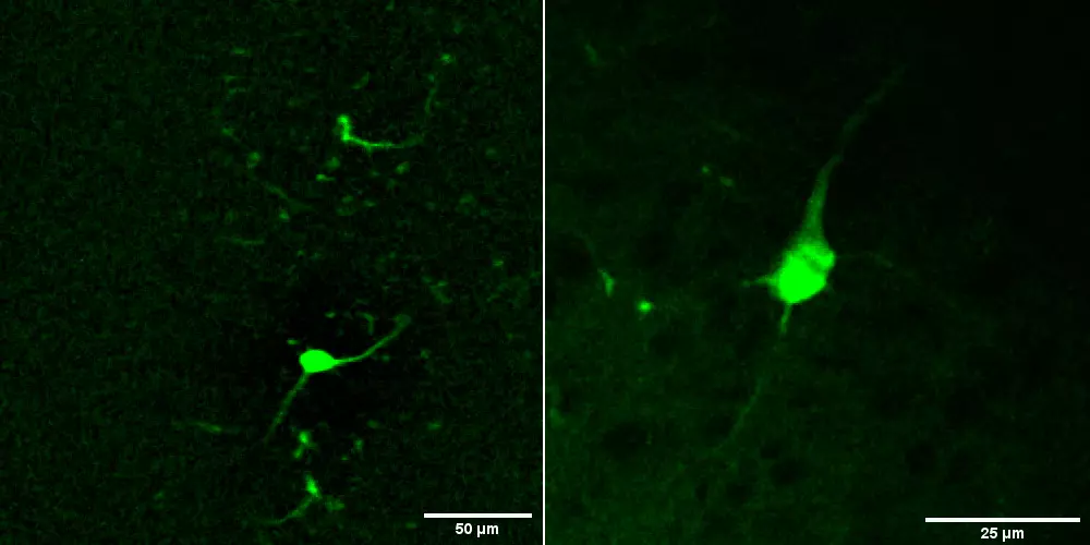
In this research line we took advantage of three-photon fluorescence microscopy (3PFM) to extend the access to neuronal activity information at deeper structures of the brain. Imaging with 3PFM will enable deeper and higher resolution imaging compared to two-photon or single-photon fluorescence microscopy. Longer excitation wavelengths reduce the tissue scattering and the higher order nonlinear excitation increases the background suppression, thus overcoming the depth limit imposed by the signal-to-background ratio of TPFM. These properties allow to achieve hippocampal CA1 functional imaging at ~1 mm depth within an intact mouse brain (Ouzounov et al., Nat Methods, 2017), or permits to perform neuronal imaging in L2/3 of mouse cortex through intact skull (Wang et al., Nat Methods 2018). Exploiting this new technology, we plan to increase our capability to study the brain structure and functions in different animal models and also to study aspects of pathological conditions otherwise hardly examined due to technological limitations.
Highlights
In the realm of modern neuroscience, the quest to unravel the complexities of the brain has led researchers to innovate continuously. One such groundbreaking area is 3P Neuronal Imaging, a…