The technique, applied for the first time to brain tissue, makes it possible to examine paraffin-embedded samples in detail and in three dimensions at the cellular level. Such samples are easy to obtain because they are very common in hospitals, and the method provides new insights into diseases such as epilepsy.
Researchers from the Departments of Biology, Physics and Astronomy, and NEUROFARBA at the University of Florence, together with the European Laboratory for Nonlinear Spectroscopy (LENS), have developed a new method that allows rehydration and analysis of human tissue samples preserved in paraffin for decades. The study, published in Communications Biology, demonstrated for the first time that this technique can examine three-dimensional samples of human brain tissue and link structural information from the cortex to developmental disorders, including epilepsy. The work was conducted at the LENS biophysics unit, directed by Francesco Saverio Pavone. The results were obtained thanks to a multicenter effort involving prestigious Italian research centers, including the Meyer Children’s Hospital, the Carlo Besta Neurological Institute, and the University of Padua, which provided the valuable samples analyzed in the study.
“We could speak of a sort of ‘brain cold case’—cases from which we have now obtained new and valuable information years later,” says Irene Costantini, a researcher at the University of Florence and lead author of the study. “These samples were collected years ago and stored in hospital biobanks, often in the hope that future technologies would allow for more detailed analyses,” she explains. Thanks to innovative methodologies, the study made it possible to use these materials to obtain new and fundamental information that has helped clarify the pathogenesis of patients’ conditions.
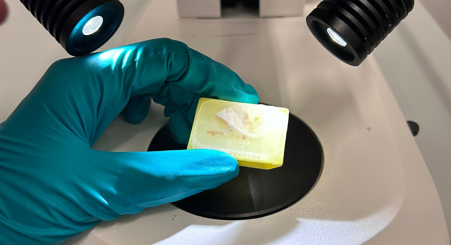
The analyzed samples had been embedded in paraffin—a material that allowed their preservation in hospital archives for decades—and were later “cleaned” for analysis. The samples used in the study date back 11 years, which is quite a long time in tissue analysis, and the researchers believe the method could be extended to even older samples.
The LENS researchers applied the method to two pathological brain samples from pediatric patients with focal cortical developmental malformations. These brain abnormalities can cause epilepsy and cognitive or motor impairments. The samples were first deparaffinized—by removing the protective coating—and then rehydrated. While this system has been used before for biological samples embedded in paraffin, this is the first time it has been successfully applied to brain tissue. The study also used a tissue-clearing technique—a method that makes biological tissue transparent and easier to observe under a microscope. Once cleaned, the tissues were examined in three dimensions using two-photon fluorescence microscopy, which provides submicrometric resolution.
Traditional histology techniques—the study of biological tissues—have intrinsic limitations, as they rely on tissue sections thin enough for microscope light to pass through, thus offering only fragmented information. “Examining a sample in three dimensions allows us to obtain much more information about its structure while preserving the original spatial context and revealing details that might be missed in two-dimensional sections,” explains Danila Di Meo, first author of the study. “It’s like the difference between reading several pages of a book and stopping at just one.”
The researchers then used machine learning techniques to analyze the volume, shape, density, and clustering of the pathological cells. “It’s a huge amount of data that requires artificial intelligence methods to process,” explains Michele Sorelli, co–first author with Di Meo. “We developed a data-processing pipeline that enables the characterization of neuronal body morphology and their three-dimensional spatial distribution.” By comparing structural arrangements with genetic information from the pathological cells, it is possible to associate specific structural characteristics with particular genetic variants. “We discovered that the two samples we analyzed—classified as the same disease but with two genetically distinct variants—actually showed different cellular shapes and spatial distributions,” says Costantini. “This is valuable information for characterizing and potentially treating the pathology.”
Moreover, obtaining biological samples of human brain tissue—especially from rare disorders—is extremely difficult. “Our understanding of the brain and its diseases is still limited by the number of samples available,” the researchers explain. “With this study, we have shown that it is possible to access a widespread archive of brain samples that can now finally be studied using modern analytical techniques.” Such an archive exists in many hospitals and could open the door to new clinical applications. “For example, in epilepsy, one-third of individuals develop drug resistance, leaving surgical removal of the epileptogenic area as the only option. With more epileptic brain samples available for analysis through this technique, we can improve both diagnosis and treatment outcomes, providing crucial information to surgeons and therapists.”
References
Di Meo, D., Sorelli, M., Ramazzotti, J. et al. Quantitative cytoarchitectural phenotyping of deparaffinized human brain tissues. Commun Biol 8, 1527 (2025). https://doi.org/10.1038/s42003-025-08887-y