A groundbreaking optical technique developed at LABS uses transparent zebrafish to reveal how brain regions communicate in real time
Understanding how different areas of the brain communicate with each other remains one of the great challenges in neuroscience. A recent study by the research group led by Francesco Saverio Pavone, published in Communications Biology, marks a major step forward in this direction. The team has developed a pioneering “all-optical” method to map the brain’s connectivity in living organisms, using larval zebrafish as a model.
By combining advanced imaging and optical stimulation, this approach allows researchers not only to observe neuronal activity in real time, but also to manipulate it with extraordinary precision. This enables scientists to explore not just how different brain areas are activated together, but how one region may causally influence another — a fundamental leap in understanding how the brain works as an integrated system.
Zebrafish and light: a powerful alliance for brain research
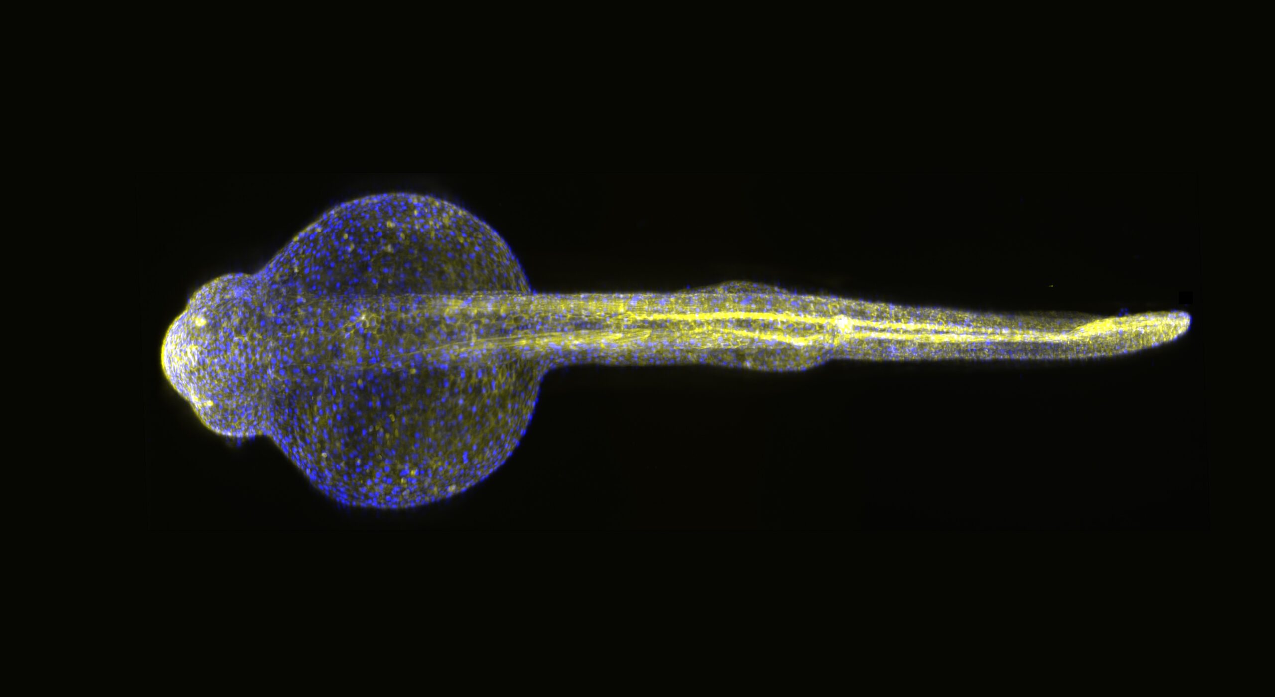
Zebrafish larvae offer a unique advantage in neuroscience: their brains are small, relatively simple, and completely transparent. This transparency makes them ideal subjects for optical imaging. Moreover, many of the basic mechanisms of brain function are conserved across species, making findings in zebrafish relevant to human neuroscience.
Pavone’s team combined two cutting-edge technologies: two-photon microscopy, which allows deep, high-resolution imaging of brain structures, and optogenetics, a technique that uses light to activate or inhibit specific neurons. The result is an “all-optical neurophysiology” platform that enables scientists to observe and influence neural circuits simultaneously, without physical electrodes or invasive procedures.
“These technologies allow us to interact with the brain in a powerful way”, explains Francesco Saverio Pavone. “By using light both to record and to stimulate neural activity, we can explore the brain’s functional architecture with unprecedented precision — all while keeping the system intact and alive. This opens up exciting possibilities for non-invasive neuroscience assisted by AI”.
Mapping the brain in action: from correlation to causality
A key innovation of the study lies in its ability to explore not just functional connectivity — statistical correlations between neuronal activities — but also effective connectivity, which refers to directional, causal relationships between brain regions.
Using their optical system, the researchers stimulated specific neurons and recorded how the rest of the brain responded. This approach allowed them to generate detailed connectivity maps of the zebrafish brain, highlighting both the regions involved and the direction of information flow. The findings offer a new perspective on how complex patterns of activity emerge from relatively simple neural architectures and open the door to exploring how these patterns might be disrupted in neurological diseases.
Looking ahead: why this research matters
The implications of this work extend far beyond the lab. All-optical neurophysiology in zebrafish provides a scalable, non-invasive, and high-resolution method to investigate the brain’s inner workings. It may contribute to a better understanding of neurodevelopmental disorders, neurodegenerative diseases, and even the neural basis of behaviour and cognition.
“Our goal is to build a deeper, mechanistic understanding of how the brain works, not just in health, but also in disease”, says Pavone. “Techniques like this bring us closer to identifying the neural signatures of complex behaviours and disorders, and could one day inform new therapeutic strategies or brain-inspired technologies”.
The research, carried out at the Laboratory for Advanced Biological Sensing (LABS) within the European Laboratory for Non-linear Spectroscopy (LENS), demonstrates the power of interdisciplinary science, blending physics, biology, and engineering to push the frontiers of neuroscience. With technologies like this, we move one step closer to decoding the brain’s language — and, perhaps one day, to healing it.