Mesoscale Functional Imaging in awake mice (head-fixed and freely moving)

Problems
During the execution of a specific task, it is essential to have a global view of the cortical neural activity in both hemispheres of the brain. It is also very useful to be able to intervene with brain stimulation protocols (via optical stimulation), to study the correlation between the functional map and the animal’s behaviour, both in head-constraint and freely behaving, such as in social interactions.
Technology
The creation of animal models that allow the correlation of neural patterns with optical signals is fundamental also in relation to transgenic models connected to All-Optical Electrophysiology techniques. We use widefield calcium imaging to investigate how different cortical areas interact with each other while mice perform skilled goal-directed motor tasks. We then study how activity across different areas of the cortex emerges during the motor output and how functional connectivity among these areas is modulated by the motor output.
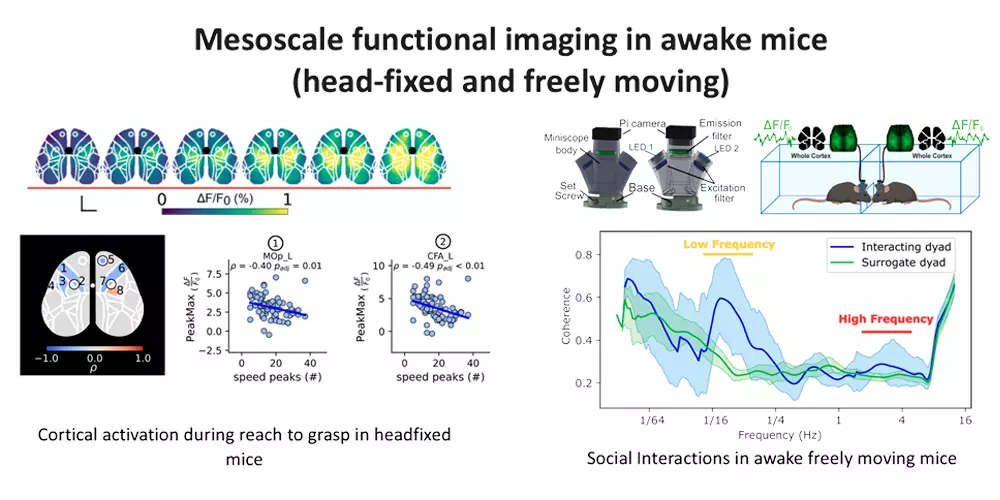
We have also developed mini microscopes, which allow us to image the neuronal signals of both hemispheres even in conditions of social interaction.
Outcomes and impact
A direct correlation between functional connectivity and brain damage or behaviour allows the dissection of the neural circuits activated during a rehabilitation phase following brain damage or to correlate their functioning with specific behaviour. This assumes particular importance when two subjects interact with each other and the detection can be done simultaneously on both by looking for possible synchronization phenomena.
More details
We are interested in how the brain processes and integrates information at the cortical level over multiple areas to produce behavioral responses and how these processes are altered in pathological conditions. To this end, we perform mesoscale functional imaging in awake mice expressing a fluorescent calcium indicator (GCaMP6f) in cortical excitatory neurons while animals are performing a behavioral task.
Cortical activity patterns during the generation of a motor output.
We use widefield calcium imaging to investigate how different cortical areas interact with each other while mice perform skilled goal-directed motor tasks. We then study how activity across different areas of the cortex emerges during the motor output and how functional connectivity among these areas is modulated by the motor output.
Cortical plasticity and reorganization in head-fixed mice after injury.
We use widefield calcium imaging to investigate cortical processing remaps in healthy mice and after a focal stroke. Daily recordings allow characterizing in the acute and in chronic phase how functional connectivity is altered after stroke and how different rehabilitative strategies act to promote cortical plasticity and functional remapping.
Developing of miniaturized device to capture cortical activity in awake freely moving mice.
We are developing small miniaturized wide-field imaging devices that allow us to break free from the head-fixed condition and allow us to study cortical activation and functional connectivity in more naturalistic behavioral settings.