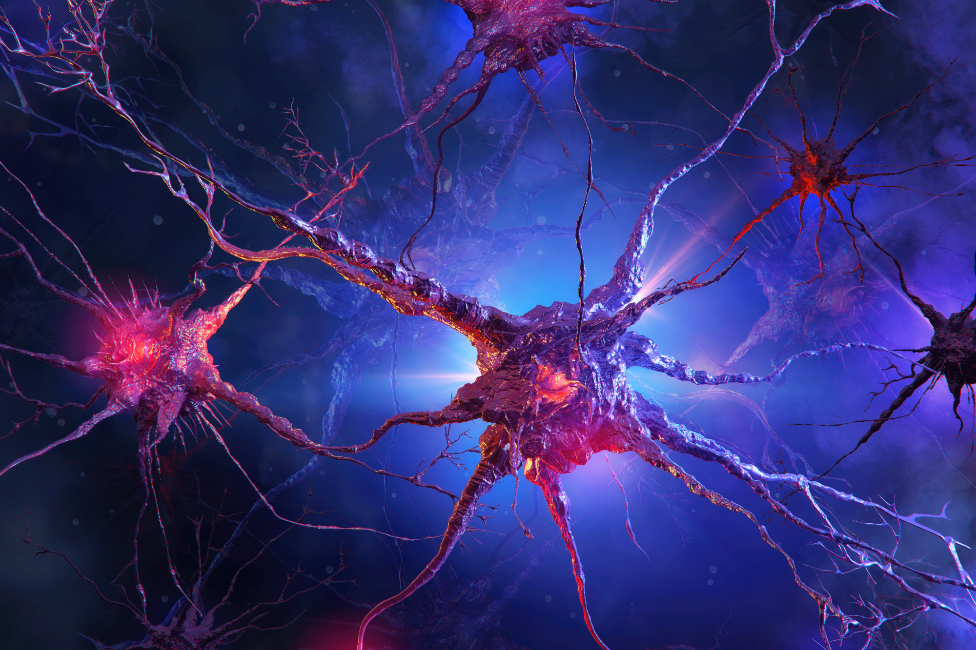
In the realm of modern neuroscience, the quest to unravel the complexities of the brain has led researchers to innovate continuously. One such groundbreaking area is 3P Neuronal Imaging, a technique that promises to revolutionize our understanding of brain function and structure. We had the privilege of sitting down with Professor Francesco Pavone, Group Leader at LABS and a pioneering figure in this field, to discuss the intricacies and implications of this cutting-edge research.
Prof. Pavone, could you explain what 3P Neuronal Imaging is and what makes it unique in the study of the brain?
3P, or three-photon microscopy, is a remarkable advancement in fluorescence imaging. Unlike traditional methods, it allows us to delve deeper into the brain tissue with minimal invasiveness. This is crucial for studying intricate neural networks and understanding how they function at both a cellular and a molecular level.
What drove the development of this technique, and how does it enhance our understanding of neurological diseases?
The need to observe the brain’s inner workings without disrupting its natural state was a key driver. With 3P Neuronal Imaging, we can study live brain tissues and observe how brain traumatic injury or stroke, tumors, or neurodegenerative diseases alter neural activity. It’s a significant leap towards early diagnosis and better treatments.
Can you share some insights on the challenges you faced in developing this technology?
One of the biggest challenges was balancing the depth of penetration with resolution, photons flux, and speed. We wanted to observe the brain as accurately and as quickly as possible, without compromising the quality of our images or damaging the sample. Achieving this required not just technological innovation, but also a deep understanding of neuroscience.
How does this method compare with traditional brain imaging techniques?
Traditional methods like MRI (Magnetic Resonance Imaging) or CT (Computed Tomography) scans offer a broader view of the brain but lack the resolution and cell specificity that 3P provides. Our method allows us to see the finer details of neuronal activity and structure, which were previously obscured.
Looking ahead, what are the potential applications of 3P Neuronal Imaging? What’s next for your research?
The applications are vast. From advancing our basic understanding of the brain to developing new therapies for neurological conditions, the potential is immense. We are now working towards making this technology more accessible and further refining it to explore even deeper aspects of the brain.