Biomedical Imaging and Spectroscopy
Problems
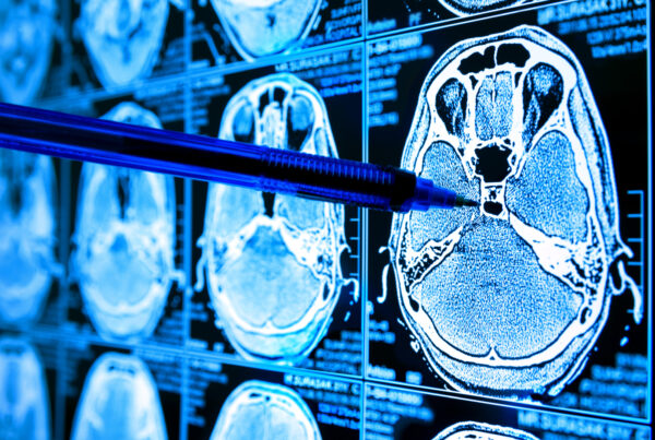
Creating a new online biopsy method (without the need to extract the tissue sample), both on biological tissues and on body fluids, allows for the diagnosis of tissue pathologies in a very early stage of the disease (tumours, neurodegenerative diseases, and all those diseases that alter the morphology of the tissue).
Technology
Here we deal with the realization of an imaging and spectroscopy systems providing information both on the morphology and on the chemical composition of the tissue. This can be done online on patients or mice, or offline on biopsies, to provide additional information to that obtained in the standard biopsy. Spectroscopic information can also be obtained from biological fluids.
Outcomes and impact
The goal is the realisation of new 3D Digital Systems for histology on biopsies. This will constitute a new online (non-invasive) biopsy systems, and new support systems for the intraoperative removal of pathological tissues. Together with this, new types of wearable sensors for online monitoring of physiological parameters will be realised.
More details
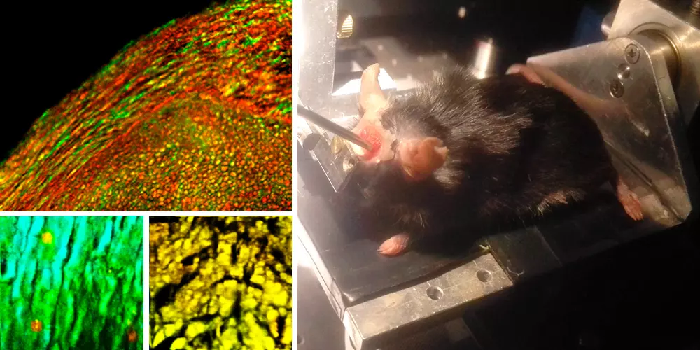
In this research line different imaging and spectroscopic methods are used to investigate the relation between morphology and molecular content in biological tissues, both in healthy and pathological condition in in-vivo and ex-vivo samples.
As a first approach we are using a multidimensional scanner comprising two and three photon excitation detection, SHG and THG, CARS and Raman imaging. This is enabling to correlate collagen structure to molecular content, cell shapes, lipids and cholesterol formation, metabolism and several others labels to understand the origin of pathology and study the reactions to therapies. This kind of morpho-functional imaging is applied both to ex vivo samples and in vivo one. Together with this, the analysis of the pathology is complemented by a pure spectroscopic analysis aiming to characterize the molecular content of the tissue. To this aim, a fiber optic probe is developed to perform multidimensional spectroscopy, ranging from one photon fluorescence, Raman scattering, and diffusive reflectance spectroscopy.
Finally, in order to extend the morphochemical analysis to 3D large sample imaging, we are developing a new paradigm of 3D digital histology by developing a large area light sheet microscope to image 3D cleared samples of pathological tissues. To this aim, a fiber optic probe has been designed for implementing multiple spectroscopic techniques within the same device. Such multimodal configuration combines one photon fluorescence, Raman scattering and diffusive reflectance spectroscopies, and fluorescence lifetime analysis.
Highlights
In the ever-evolving landscape of medical science, the ability to see beyond the visible has always been a pursuit of researchers and clinicians alike. The field of Biomedical Imaging and…