Join LABS in advancing neuroscience: Develop software for 3D image analysis in fluorescence microscopy. Apply now for a role in groundbreaking brain research.
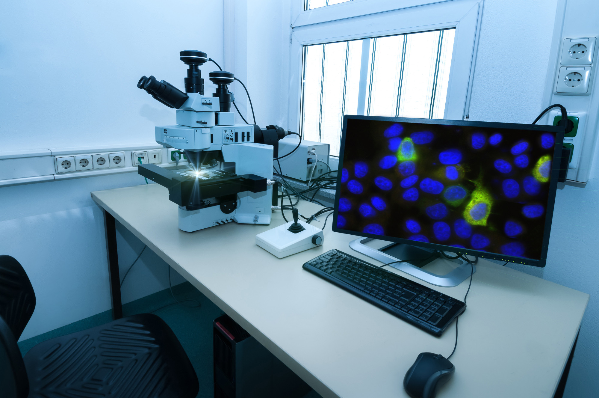
Fluorescence microscopy allows the visualization of brain structures at a micrometric resolution of volumetric samples allowing the investigation of the cytoarchitecture of biological samples previously cleared and treated with specific immunofluorescence techniques. Automatic analysis methods of the acquired images, such as two-photon microscopy and light sheet microscopy, allow the evaluation of the spatial orientation of the neuronal fibres and the cell counting in 3D. The candidate will be in charge of optimizing and developing software to analyze this type of image: from the post-processing of the individual stacks to obtain the complete fused volume of the 3D reconstruction, to the actual analysis for the extrapolation and quantification of the data of interest. The candidate will also have to organize and analyze experimental data using statistical methods and optimize procedures to allow the analysis of the large amount of data (TB) produced. Finally, the candidate will have to apply the developed methods to the volumetric reconstructions of an entire human brainstem obtained with the above-mentioned fluorescence microscopies, to evaluate the density and 3D organization of the fibres and cells present in the biological tissue.
The deadline for submitting the application for the selections is 11/01/2024.
If you have any questions please write to pavone@lens.unifi.it


